Q) False regarding the management of Acute Diverticulitis, Sigmoid colon inflammation and Fat stranding in CT ?
a) Outpatient treatment in most cases
b) Do a colonoscopy after the resolution of acute symptoms
c) Elective Colectomy to be done
d) IV antibiotics to be started`
The pathogenesis of acute diverticulitis is often attributed to the obstruction of diverticula by fecaliths, leading to increased intraluminal pressure, bacterial overgrowth, and subsequent inflammation or perforation
Answer Free c
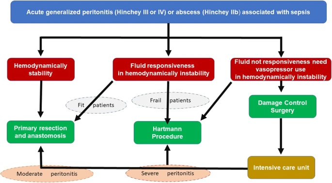
Sigmoid diverticulitis can be complicated and uncomplicated
Complicated means diverticulum associated with abscess, perforation, obstruction, fistula
This question is about an uncomplicated acute diverticulitis
It can be managed in outpatient setting
It requires IV antibiotics and diet modification
After resolution of symptoms, colonoscopy is to be done after 6 weeks to rule out the presence of other diverticula and neoplasm
Colectomy is not required in all cases. Current recommendations suggest that the decision for surgery should be individualized, taking into consideration the frequency and severity of recurrences. The patient’s overall medical condition and comorbidities should also be included in the analysis
Q) A 60 year old male is contemplating hyperbaric oxygen therapy for radiation proctitis. Which of the following is not true regarding this?
A. Indicated in acute radiation proctitis but not in subacute or chronic radiation proctitis B. Oxygen increases the growth of residual tumor and hence tumor should be completely resected C. Complications include Parkinsonism, barotrauma D. Usually 30-40 sessions are required for treatment
Ans a
This statement is not true because hyperbaric oxygen therapy (HBOT) is not only indicated in acute radiation proctitis but can also be beneficial in subacute and chronic radiation proctitis. Studies have shown that HBOT can promote healing in chronic radiation injuries as well.
Hyperbaric oxygen overcomes chronic tissue hypoxia in radiation damaged tissues and with repeated sessions induces growth of regenerative tissue, capillaries, and epithelium. Successful therapy may take multiple sessions. 18 to 60 treatments
HBO treatments for hypoxic wounds are usually delivered at 1.9 to 2.5 atm for sessions of 90 to 120 minutes each. Treatments are given once daily, five to six times per week and should be given as an adjunct to surgical or medical therapies. Clinical evidence of wound improvement should be noted after 15 to 20 treatments.
Complications of HBO therapy are caused by changes in atmospheric pressure and elevated PO2. Middle ear barotrauma, ranging from tympanic membrane hyperemia to eardrum perforation, is the most common complication.
Pneumothorax brain oxygen toxicity, manifested by convulsions resembling grand mal seizures; oxygen lung toxicity, resulting from damage from oxygen free radicals to lung parenchyma and airways and ranging from tracheobronchitis to full-blown respiratory distress syndrome; and transient myopia.
Absolute contraindications to HBO therapy are
(1) uncontrolled pneumothorax
(2) current or recent treatment with bleomycin or doxorubicin (potential aggravation of cardiac and pulmonary toxicity), and
(3) treatment with disulfiram (increases risk of developing oxygen toxicity).
a) The optimal treatment for lymphomas unresponsive to initial H. pylori antibiotic treatment remains unclear and includes the chemotherapy, radiotherapy, surgical resection, etc
b) Almost all MALT-lymphoma may regress with conventional H. pylori treatment.
c) Need for surgery in lymphoma is mainly for its complication
d) Risk of perforation is over estimated in the literature
Q. All are true about adenomatous polyposis syndrome except? ( Repeated in NOV INI SS) A. 25% do not have family history B. Attenuated FAP has less than 100 polyps and delayed onset (50-55 yrs) C. More than 20 rectal polyps have to be operated as there is high risk of Carcinoma D. Attenuated FAP don’t have extracolonic manifestations and carry APC mutation
Ans d
Attenuated FAP is a phenotypically distinct variant of FAP in which
(1) affected individuals have fewer than 100 adenomas
(2) the polyps are more proximally distributed in the colon, and
(3) the onset of colorectal cancer is about 15 years later than in patients with FAP.
Germline mutations in the APC gene are found in 80% to 90% of patients with classic FAP and in 10% to 30% of patients with AFAP.
About 25% of patients with FAP have a de novo mutation and thus have no family history.
Attenuated FAP is a milder form of classic familial adenomatous polyposis (FAP) and is characterized by fewer colon polyps (an average of 30) and a delay in the development of colon cancer (average age 50 to 55 years) (True)
AFAP is caused by mutations in the APC gene and is inherited in an autosomal dominant manner.
Mutations responsible for this variant occur in the extreme upstream or downstream
portions of the APC gene.
Q) Antral GIST 1cm incidentally found on UGIE. True regarding its management a) Surgical resection b) Endoscopic resection c) Resection required if EUS suggests irregular border with cystic spaces d) Endoscopic surveillance, if size >2cm then resect
Ans
c
GIST more than 2 cm should undergo resection. Management of GIST less than 2 cm is dependent on weather high risk features are present on EUS.
The high risk features are
1. Irrgeular margins
2. Heterogenous architecture
3. Ulcers
Management of low risk GIST less than 2 cm is surveillance every 6 months
Q . Treatment of choice for anal canal high grade lymphoma on a young immunocompromised male? A. Chemoradiation B. APR C. Local excision followed by chemotherapy D. Local excision
Q) All are true about pancreatic protocol CT except (#AIIMS )
a) > 90% of un resect able lesions are picked up by CT
b) It is a dual phase CT with cuts taken at 40 secs and 70 secs
c) Liver metastasis are detected in early arterial phase
d) All are true
Ans c
Pancreatic protocol CT involves imaging at the pancreatic phase (i.e., approximately 45 seconds after contrast administration) and at the portal venous phase (i.e., approximately 70 seconds after contrast administration).
It is useful for detection of adenocarcinoma of pancreas.
Metastatic lesions are seen in the portal venous phase, because the lesions are not typically well vascularized.
Arterial phase images are principally used to distinguish metastatic disease from benign vascular lesions, such as hemangiomas, or to better define the arterial anatomy of the liver.
Non contrast phase used for
Evaluation of pancreatic calcifications and allows localization of the precise levels for imaging on the post contrast study.
Early arterial phase
Evaluation of pancreatic vasculature without interference from venous opacification.
Late Arterial Phase
Distinguish pancreatic neoplasms from adjacent normal pancreatic tissue It also is useful to evaluate hypervascular liver metastases as seen in patients with neuroendocrine tumors of the pancreas.
Portal Phase
Evaluate for hypovascular liver metastases
Ref Blumgart
The dedicated pancreas protocol uses 750 to 1000 mL of oral water as a negative contrast agent administered before the examination, to aid distinction of enhanced vessels from the
gastrointestinal tract