During anorectal manometry, a patient demonstrates preservation of resting continence despite severe internal anal sphincter damage. Which structure is most responsible for maintaining continence in this scenario?
A. Pubococcygeus
B. Iliococcygeus
C. Puborectalis
D. External anal sphincter
Answer: C. Puborectalis
Explanation:
The puborectalis muscle forms a U-shaped sling around the anorectal junction and is the most critical muscle for maintaining fecal continence. Its tonic contraction maintains an acute anorectal angle, preventing involuntary stool passage. Even with internal anal sphincter damage, the puborectalis sling can preserve continence.
Teaching Points:
Puborectalis forms a sling at the anorectal junction.
Maintains anorectal angle (~80–90°) → continence.
Relaxes during defecation → angle straightens.
Dysfunction → dyssynergic defecation or fecal incontinence.
Key compensatory mechanism when sphincters are weak.
Clinical Scenario: A 52-year-old hypertensive male presents with sudden retrosternal chest pain radiating to the back. CT angiography reveals dissection confined to the ascending aorta without involvement of the arch or descending thoracic aorta.
Which DeBakey classification does this correspond to?
A. Type I
B. Type II
C. Type IIIa
D. Type IIIb
Correct Answer: B. Type II
Explanation:
Type II: Limited only to ascending aorta
Requires emergency surgical repair → risk of tamponade
Type I: Extends from ascending → arch → descending
Type III: Origin distal to left subclavian; involves descending aorta
Quick Memory Tip:
“Type II = A = Ascending only → Always surgical”
Q) A 60-year-old patient undergoes EUS for staging of an early esophageal tumor. Regarding the echogenic layers of the esophageal wall, which of the following statements is INCORRECT?
✅ Answer: c) The fourth hyperechoic layer represents the muscularis propria (incorrect — the fourth layer is hypoechoic and corresponds to muscularis propria).
🔍 Explanation:
EUS shows 5 layers of the esophageal wall:
Correct identification is critical for T-staging of esophageal cancer. Misidentifying layer 4 may lead to incorrect staging and management errors. Radial echoendoscopes provide optimal visualization of all layers.
Q) A 28-year-old male is brought to the ED after a road traffic accident with polytrauma. He undergoes emergency laparotomy for splenic injury. On postoperative day 1, he develops fever (38.7°C), tachycardia (120/min), leukocytosis (18,000/µL), and hypotension requiring fluids. Blood and urine cultures are negative. No evidence of pneumonia is seen on chest X-ray.
Which of the following best explains his condition?
✅ Answer: B. Sterile systemic inflammatory response due to DAMP release
🔍 Explanation: Trauma and major surgery cause tissue necrosis, ischemia, and cellular injury. Intracellular molecules such as HMGB1, mitochondrial DNA, ATP, uric acid, and heat shock proteins are released and act as DAMPs (damage-associated molecular patterns).
These activate innate immune receptors like Toll-like receptors and inflammasomes (e.g., NLRP3), triggering a robust inflammatory response even in the absence of infection. This explains sterile SIRS, which can mimic sepsis but with negative cultures.
🧠 Key Point: DAMP-driven sterile inflammation is common after trauma, burns, pancreatitis, and ischemia-reperfusion injuries. It must be differentiated from infection-driven SIRS (PAMP-mediated sepsis).
Q) A 42-year-old male presents with multiple recurrent duodenal ulcers, abdominal pain, and chronic diarrhea. Fasting serum gastrin levels are >1000 pg/mL. Which of the following is the most likely ulcerogenic cause of hypergastrinemia?
✅ Answer: B. Zollinger–Ellison syndrome
🔍 Explanation:
Zollinger–Ellison syndrome (ZES) is caused by a gastrinoma (a gastrin-secreting tumor), typically located in the pancreas or duodenum.
It leads to massive hypergastrinemia, increased gastric acid secretion, and multiple, recurrent, or atypical peptic ulcers.
Diarrhea and steatorrhea are common due to acid inactivation of pancreatic enzymes.
Other Options:
A. Atrophic gastritis:
Leads to hypochlorhydria/achlorhydria with secondary hypergastrinemia, but non-ulcerogenic (low acid state).
C. Chronic PPI use:
Causes compensatory hypergastrinemia due to acid suppression, but again non-ulcerogenic unless stopped abruptly in predisposed individuals.
D. Helicobacter pylori infection:
May increase gastrin levels mildly, but ulcers are primarily due to mucosal damage and inflammation, not from gastrin hypersecretion.
🧠 Key Point: Zollinger–Ellison syndrome is the only ulcerogenic cause of hypergastrinemia. Fasting gastrin >1000 pg/mL with low gastric pH is diagnostic.
Q: A 30-year-old male presents to the emergency department after a stab wound to the right chest.
On examination, there is a 4 cm open wound in the 5th intercostal space anteriorly, with a sucking sound during inspiration, decreased breath sounds on the right, and respiratory distress.
What is the next best step in management?
# Theme NEET SS Mocktest 1
A) Immediately close the wound with an airtight dressing
B) Insert a chest tube on the same side and then close the wound
C) Intubate and initiate positive pressure ventilation
D) Apply a three-sided occlusive dressing to the wound
🆓 This is a free MCQ — click below to view the answer.
✅ Correct Answer: D) Apply a three-sided occlusive dressing to the wound
Explanation: This is a classic open pneumothorax (sucking chest wound). When the defect is ≥2–3 cm, air enters the pleural cavity preferentially, impairing ventilation.
Management:
- First: Apply a three-sided occlusive dressing. This allows air to escape on exhalation but prevents entry during inhalation, avoiding tension pneumothorax.
- Then: Insert a chest tube on the same side before sealing the wound completely.
Why others are incorrect:
A) Airtight closure before chest tube risks tension pneumothorax.
B) Chest tube insertion is essential but follows the temporary dressing.
C) Intubation may be required later, but immediate chest wound control is priority.
Q: During a surgical skills assessment, you are asked to perform a hand-sewn intestinal anastomosis using a continuous, inverting suture that enters the bowel lumen.
Which of the following suture techniques best fits this description?
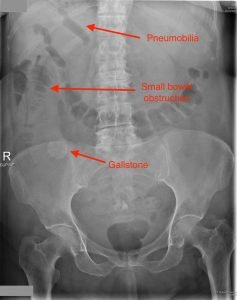
A 65-year-old male presents with abdominal pain, vomiting, and a history of multiple episodes of cholecystitis. X ray image is given below.
What is the most likely diagnosis?
A. Acute cholecystitis B. Gallstone ileus C. Small bowel volvulus D. Duodenal perforation
Answer:B. Gallstone ileus
Explanation: Rigler's Triad consists of pneumobilia, small bowel obstruction, and an ectopic gallstone, which is diagnostic of gallstone ileus. This condition occurs when a gallstone enters the bowel through a biliary-enteric fistula, leading to mechanical obstruction.
A large gallstone (>2.5 cm) erodes through the gallbladder wall, creating a cholecysto-enteric fistula (most commonly into the duodenum).
The stone enters the bowel and may cause obstruction, most often at the ileocecal valve due to its narrow lumen.
The presence of air in the biliary tree (pneumobilia) results from communication between the biliary and intestinal tracts.
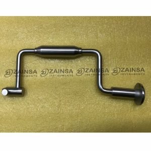
The Hudson brace is a manually operated surgical drill used in neurosurgery and orthopedic procedures. It consists of a hand-cranked mechanism with interchangeable drill bits for trephination or skull perforation.
Q: Which of the following statements is most accurate regarding axillary lymph node dissection (ALND) in breast cancer staging?
a) Level I and level II ALND requires the removal of at least 10 lymph nodes for accurate staging, and level III nodes should always be included in the dissection, regardless of the presence of gross disease in levels I and II.
b) The axillary dissection should include tissue from levels I and II, with a focus on the area inferior to the axillary vein, extending laterally to the latissimus dorsi muscle and medially to the pectoralis minor muscle, when there is no gross disease in level II nodes.
c) Level III nodes should be dissected in all cases of breast cancer for accurate staging, as they are always involved in metastatic spread.
d) Level I and level II ALND can be skipped in cases of clinically negative axilla, as there is no need for lymph node evaluation in the absence of suspicion of metastasis.
🔒 This is a premium MCQ. Only logged-in premium members can view the answer.