Right gastroepiploic vein

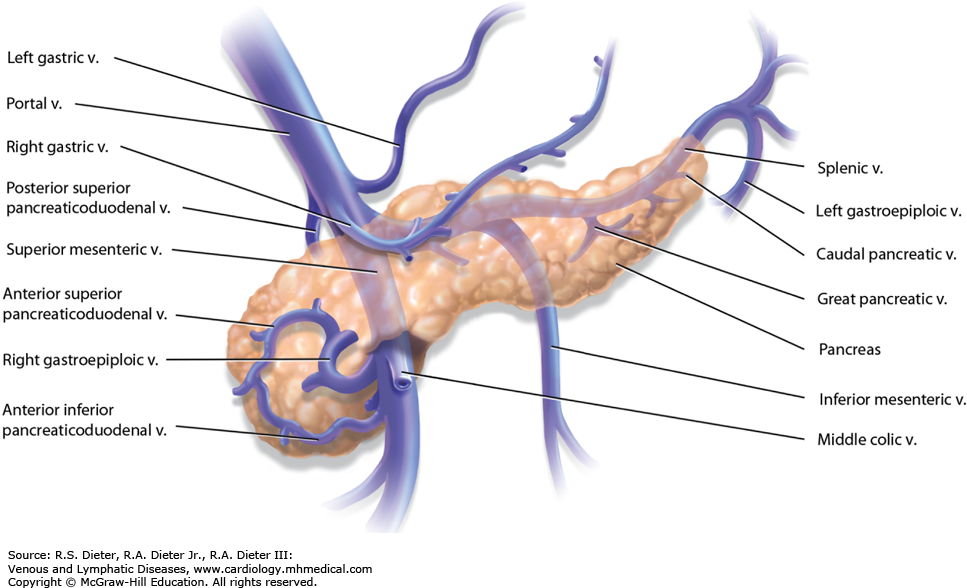
Q) Right gastroepiploic vein drains into
A. Splenic vein
B. Left gastric vein
C. Portal vein
D. Superior mesenteric vein


ANswer is free

D

SMV

Veins of SMV
Right gastro epiploic vein

The right gastroepiploic vein is a significant blood vessel located in the abdomen. It runs parallel to the right gastroepiploic artery and is an essential part of the venous drainage system of the stomach.

Originating from the greater curvature of the stomach, this vein receives blood from various branches, including the short gastric veins. As it continues its course, it eventually joins with the superior mesenteric vein, contributing to the portal venous system. Understanding the anatomy and function of the right gastroepiploic vein is crucial for medical professionals in diagnosing and treating related conditions, ensuring proper circulation and overall digestive health.

The right gastroepiploic vein is essential in many surgical procedures:

  • Gastric Bypass and Gastric Cancer Surgery: The RGEV may require ligation or resection in gastrectomy procedures. Surgeons should be cautious of potential bleeding risks and the implications for vascular flow.
  • Coronary Artery Bypass Grafting (CABG): Surgeons often use the right gastroepiploic artery as a graft, though the associated vein is also considered during CABG preparation due to its proximity and importance.
  • Trauma and Emergency Surgery: Understanding Right gastroepiploic vein anatomy aids in identifying bleeding sources, especially in abdominal injuries.

Questions on Liver 

Surgery Anatomy

Pseudoachalasia

Q) Most common cause of  pseudoachalasia is ?

(a) Benign tumors of esophagus

(b) Chagas disease

(c) Caustic injury

(d) Adenocarcinoma of cardia


d

Pseudoachalasia is an achalasia-like disorder that is usually produced by adenocarcinoma of the cardia

Other uncommon causes are

 1.benign tumours at this level.

2, Tumors of bronchus, pancreas

It  is a condition that mimics the symptoms of achalasia, but is caused by a different underlying problem. The most common cause  is  malignancy in the gastroesophageal junction (GEJ), which is the area where the esophagus meets the stomach. Other possible causes of pseudoachalasia include:

  • Esophageal stricture
  • Chagas disease
  • Radiation therapy to the chest
  • Aortic aneurysm
  • Thyroid cancer

Pseudoachalasia presents in an identical manner to idiopathic achalasia with progressive dysphagia to solids and liquids, retrosternal pain, regurgitation of undigested foods and weight loss.

The main difference between pseudoachalasia and achalasia is that it  is often associated with other symptoms, such as abdominal pain, vomiting, and weight loss.

The diagnosis is made through a combination of clinical evaluation, upper endoscopy, and esophageal manometry. Upper endoscopy can help to rule out a malignancy in the GEJ, and esophageal manometry can help to confirm the diagnosis of achalasia. In some cases, a CT scan or MRI of the chest may be needed to further evaluate the cause of pseudoachalasia.

Pseudoachalasia

Questions MCQs on Esophagus 

Case report 

Marjolin Ulcer

Q) 35 year old male has come with Marjolin ulcer if the leg. True about Marjolin's ulcer is ?

a) Lymphatic spread is common

b) They are painful

c) Aggressive and fast growing tumors

d) Squamous cell carcinoma is the most common type


d. squamous cell carcinoma

When a SCC or BCC occurs in a long standing scar, it is called marjolin's ulcer.

Marjolin's type of ulcer is a malignant change that can occur in any long standing ulcer (ie venous ulcer)

Scar tissue is devoid of lymphatics, so no lymphatic spread. Lymphatic spread can still occur when it invades normal tissue. also nerve endings are not in scar tissue, so pain is a late feature

They are slow growing tumors but have propensity for distal metastasis  and squamous cell carcinoma is the most common type. Slow growth is again due to avascular characteristic

Bailey 28th 625

Squamous cell cancer of upper esophagus

Q) A 65-year-old male presents with grade IV dysphagia and is diagnosed with squamous cell carcinoma of the upper third of the esophagus. What is the most appropriate next step in management?

a) Definitive chemoradiotherapy
b) Neoadjuvant chemotherapy followed by transhiatal esophagectomy
c) Systemic chemotherapy alone
d) Neoadjuvant chemoradiotherapy followed by three-field esophagectomy

Correct answer: a) Definitive chemoradiotherapy

💡 Explanation:

  • Upper third esophageal squamous cell carcinoma poses a challenge for surgical resection due to its proximity to the pharynx and larynx.

  • In resectable upper esophageal SCC, especially in older patients or when the tumor is very proximal, definitive chemoradiotherapy (CRT) is often the preferred treatment to avoid morbid surgery like laryngopharyngoesophagectomy.

  • Multiple guidelines, including NCCN and ESMO, recommend definitive CRT for upper esophageal SCC unless there’s a compelling reason for surgery.


Why other options are incorrect:

  • b) Neoadjuvant chemo + THE:
      THE (transhiatal esophagectomy) is not suitable for upper esophageal tumors. It doesn't provide good access to cervical/upper thoracic esophagus.

  • c) Chemotherapy alone:
      Not standard. Chemotherapy without radiation is inadequate for curative intent in localized esophageal cancer.

  • d) Neoadjuvant CRT + three-field esophagectomy:
      Though this is an option for mid/lower third esophageal cancers, especially in younger patients, it's more morbid and rarely used for upper third SCC in older patients.

More MCQS

More Esophagus MCQS

Mock tests 

Question Bank

How to be a Premium Member