Risk factors for Ca gall bladder

Q) Risk factor for CA GB are all except
A. Multiple polyp
B large gall stone >3 cm
C PSC
D pigment stone same risk as cholesterol stone

Ans visible for premium members


MCN

MCQ on Mucinous Cystic Neoplasm (MCN) of Pancreas

Q: All about MCN of pancreas are true except?

# Theme Pancreas MCQ

1. Presence of eggshell calcification in CT is suggestive of malignancy
2. Cyst fluid analysis can diagnose accurately in 80%
3. Invasive MCN is very aggressive with 30% 5YR Survival compared to adeno carcinoma
4. If MCN is non invasive, surgery is curative
🔒 Premium Members Only — Login to View Answer & Explanation
🚫 This answer is available only to Premium Members. Join Premium Surgery Course to access detailed explanations.

GIST

Q) True statement regarding GIST is  (AIIMS 2019)

a) 80% of GIST arise from stomach

b) ILeal GIST is resistant to Imatinab

c) Leiomyosarcomas do not express CD 117

d) Prognosis of GIST does not depend on the site of lesion 

Answer is in the button below and can be seen only when you are a premium member and logged in

[


c
 They can appear anywhere within the GI tract, although they are usually found in the stomach (40% to 60%), small intestine (30%), and colon (15%).
 Development of imatinib mesylate has significantly altered previous treatment strategies. Imatinib mesylate is a tyrosine kinase inhibitor that blocks the unregulated mutant c-kit tyrosine kinase and inhibits the BCR-ABL and PDGF tyrosine kinases.
Current guidelines suggest that patients with high-risk disease should receive 3 years of adjuvant  therapy at all sites
Sabiston page 1280
Ileal GIST are more malignant than stomach GIST

Leiomyosarcomas

Among the gastric tumors, there were no examples of true leiomyosarcomas, whereas there were four small intestinal, four colonic, and two rectal tumors that histologically showed features of differentiated smooth muscle cells with blunt-ended nuclei and eosinophilic, sometimes granular, cytoplasm. These tumors were, by definition, all positive for SMA, and seven also were positive for desmin. Although all leiomyosarcomas were generally negative for CD117, scattered large neoplastic spindle cells (less than 1% of tumor cells) in two intestinal leiomyosarcomas showed strong cytoplasmic positivity; these four tumors were negative for CD34.

Ref https://www.nature.com/articles/3880210

Right gastroepiploic vein

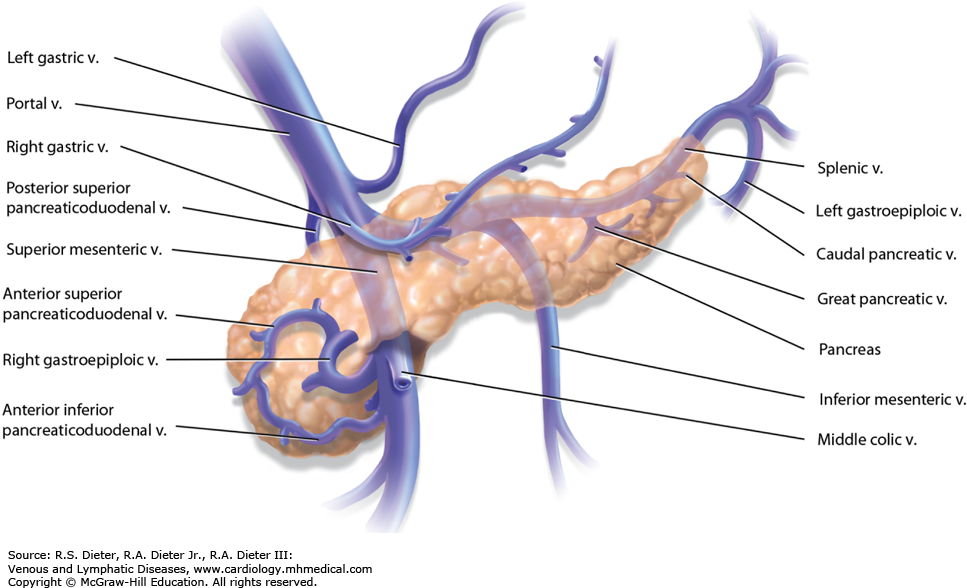
Q) Right gastroepiploic vein drains into
A. Splenic vein
B. Left gastric vein
C. Portal vein
D. Superior mesenteric vein


ANswer is free

D

SMV

Veins of SMV
Right gastro epiploic vein

The right gastroepiploic vein is a significant blood vessel located in the abdomen. It runs parallel to the right gastroepiploic artery and is an essential part of the venous drainage system of the stomach.

Originating from the greater curvature of the stomach, this vein receives blood from various branches, including the short gastric veins. As it continues its course, it eventually joins with the superior mesenteric vein, contributing to the portal venous system. Understanding the anatomy and function of the right gastroepiploic vein is crucial for medical professionals in diagnosing and treating related conditions, ensuring proper circulation and overall digestive health.

The right gastroepiploic vein is essential in many surgical procedures:

  • Gastric Bypass and Gastric Cancer Surgery: The RGEV may require ligation or resection in gastrectomy procedures. Surgeons should be cautious of potential bleeding risks and the implications for vascular flow.
  • Coronary Artery Bypass Grafting (CABG): Surgeons often use the right gastroepiploic artery as a graft, though the associated vein is also considered during CABG preparation due to its proximity and importance.
  • Trauma and Emergency Surgery: Understanding Right gastroepiploic vein anatomy aids in identifying bleeding sources, especially in abdominal injuries.

Questions on Liver 

Surgery Anatomy

Pseudoachalasia

Q) Most common cause of  pseudoachalasia is ?

(a) Benign tumors of esophagus

(b) Chagas disease

(c) Caustic injury

(d) Adenocarcinoma of cardia


d

Pseudoachalasia is an achalasia-like disorder that is usually produced by adenocarcinoma of the cardia

Other uncommon causes are

 1.benign tumours at this level.

2, Tumors of bronchus, pancreas

It  is a condition that mimics the symptoms of achalasia, but is caused by a different underlying problem. The most common cause  is  malignancy in the gastroesophageal junction (GEJ), which is the area where the esophagus meets the stomach. Other possible causes of pseudoachalasia include:

  • Esophageal stricture
  • Chagas disease
  • Radiation therapy to the chest
  • Aortic aneurysm
  • Thyroid cancer

Pseudoachalasia presents in an identical manner to idiopathic achalasia with progressive dysphagia to solids and liquids, retrosternal pain, regurgitation of undigested foods and weight loss.

The main difference between pseudoachalasia and achalasia is that it  is often associated with other symptoms, such as abdominal pain, vomiting, and weight loss.

The diagnosis is made through a combination of clinical evaluation, upper endoscopy, and esophageal manometry. Upper endoscopy can help to rule out a malignancy in the GEJ, and esophageal manometry can help to confirm the diagnosis of achalasia. In some cases, a CT scan or MRI of the chest may be needed to further evaluate the cause of pseudoachalasia.

Pseudoachalasia

Questions MCQs on Esophagus 

Case report 

Marjolin Ulcer

Q) 35 year old male has come with Marjolin ulcer if the leg. True about Marjolin's ulcer is ?

a) Lymphatic spread is common

b) They are painful

c) Aggressive and fast growing tumors

d) Squamous cell carcinoma is the most common type


d. squamous cell carcinoma

When a SCC or BCC occurs in a long standing scar, it is called marjolin's ulcer.

Marjolin's type of ulcer is a malignant change that can occur in any long standing ulcer (ie venous ulcer)

Scar tissue is devoid of lymphatics, so no lymphatic spread. Lymphatic spread can still occur when it invades normal tissue. also nerve endings are not in scar tissue, so pain is a late feature

They are slow growing tumors but have propensity for distal metastasis  and squamous cell carcinoma is the most common type. Slow growth is again due to avascular characteristic

Bailey 28th 625